Information for Providers
Medical imaging is all we do here at University Medical Imaging, which means your patients’ scans are always our top priority.
Imaging Services
When it comes to MRI, strength matters.
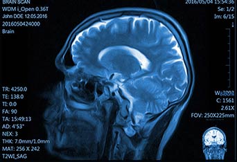
The strength of the magnetic field in MRI is measured in units referred to as Tesla (T). University Medical Imaging uses a 3.0T HD GE SIgna scanner. 3.0T is the most powerful MRI scanner currently available for clinical use. Higher-strength magnets enable improved spatial resolution contrast-to-noise (SNR), allowing detailed exploration of more complex phenomena not clearly depicted on widely used 1.5T systems.
3.0T studies of the brain, spine, chest, abdomen, pelvis, extremities, and vascular system are consistently higher in quality than those images obtained at 1.5T, providing exceptional advantages for neurology, orthopedics, vascular surgery and oncology. Bone structure, cartilage, tendons and ligaments can be clearly visualized, and pathology can be more easily detected due to improved image quality. Increased special resolution improves the capacity to image smaller structures, such as the inner ear, brachial plexus and biliary system.
Our system also helps reduce the need for contrast agents. Inhance software virtually eliminates the need for gadolinium injections in vascular imaging – a critical advantage for patients with diabetes or other kidney-related concerns.
Our robust technology also allows us to offer diffusion tensor imaging (DTI) with 3D tractography and CartiGram cartilage mapping.
For all MRI scan applications, better images mean a better report and a better diagnosis.
University Medical Imaging also offers digital X-ray and arthrogram procedures.
Are you a provider? CONNECT WITH US!
Access Images
Our referring physicians have secure, HIPAA-compliant online access to their patients' images. If you are already a registered user, click below to log in to our physician portal. If you are a referring medical professional needing access to this site, please contact us by email or at 916-922-6747 during business hours.
Go to Site